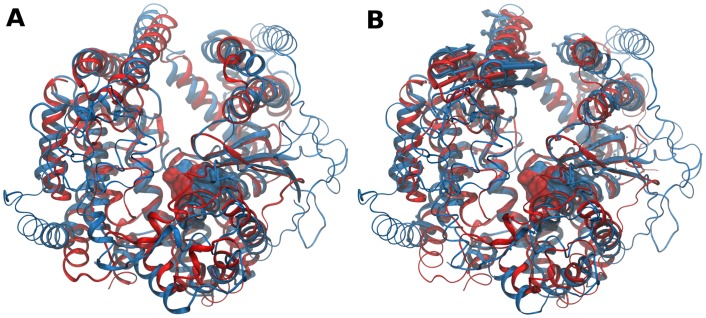
Fig 4. Structure- and dynamics-based alignments obtained for pair M3-M32.
(A) Structure-based and (B) Dynamics-based alignment of M3 representative Neurolysin (blue, PDB ID: 1I1I) and M32 representative Carboxypeptidase Pfu (red, PDB ID: 1KA4). Produced alignments were obtained using the DaliLite and ALADYN web-servers (see Methods). Aligned residues colored in cartoon representation, non-aligned residues in colored ribbons and active site residues in surface representations (Neurolysin: H474, E475, H478 and E503; Carboxypeptidase Pfu H269, E270, H273 and E299). Colored arrows indicate modes of motion of aligned portions along the first mode.

