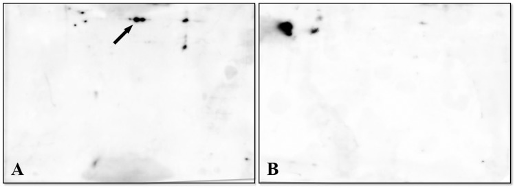
Fig 4. Immunoblotting of proteins from SG and OT profiles generated as indicated in Fig 1.
PVDF membranes were incubated with the rabbit polyclonal antibodies anti-FliD of M. mitochondrii, followed by anti-rabbit antibody. The protein spot(s) indicated by an arrow in Panel A (OT pool) was tentatively assigned to FliD. Panel B shows the SG profile in which the hypothetical FliD spot is undetectable.

