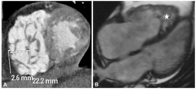
Fig. 1. A 72-year old male with noncompaction of right ventricle. A: short axis view of the ventricles on computed tomography imaging showing deep intertrabecular recesses within the right ventricle with a noncompacted to compacted myocardium ratio of 8.5. B: true fast imaging with steady-state precession (true-FISP) cine four-chamber magnetic resonance image delineating noncompaction in the apical region of the right ventricle (star).

