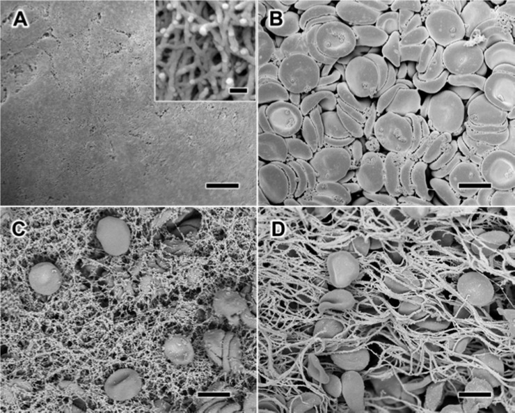
Figure 1.
Scanning electron microscopy (SEM) visualization of the morphology of the interaction between whole blood and RADA16-I. When alone in solution, RADA16-I spontaneously self-assembles into interpenetrating nanofibers (A), while anticoagulated whole blood shows the presence of red blood cells and platelets without fibrin formation (B). Mixture of RADA16-I with anticoagulated whole blood reveals the physical entrapment of blood components in a network of peptide nanofibers (C), which appears similar to naturally coagulating fibrin-based clots (D). Scale bars represent 5 μm (A–D) and 100 nm (A, inset).

