
Figure 3.
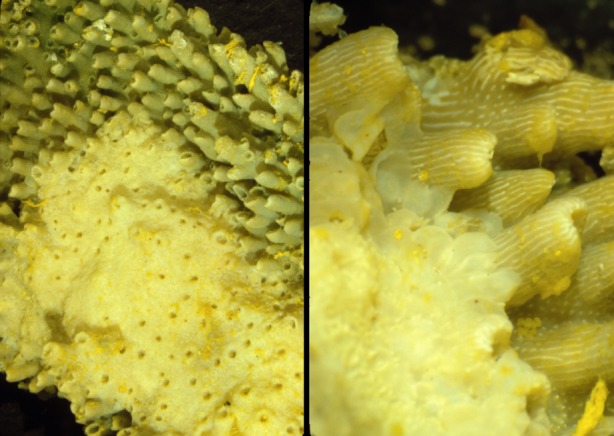
Cancer in corals. Corals often exhibit tumours called calicoblastic epitheliomas with loss of differentiation and destruction of the tissue architecture, including the mechanisms for resource allocation [113]. The normal tubular growth pattern in the upper right of both panels is being invaded by the relatively smooth, unstructured calicoblastic epithelioma. These samples are from the Grecian Rocks, Florida Keys (Florida Keys National Marine Sanctuary). The coral appears yellow because the samples were preserved in Helly's fixative which included potassium dichromate. Pictures courtesy of Esther Peters.