
Figure 1.
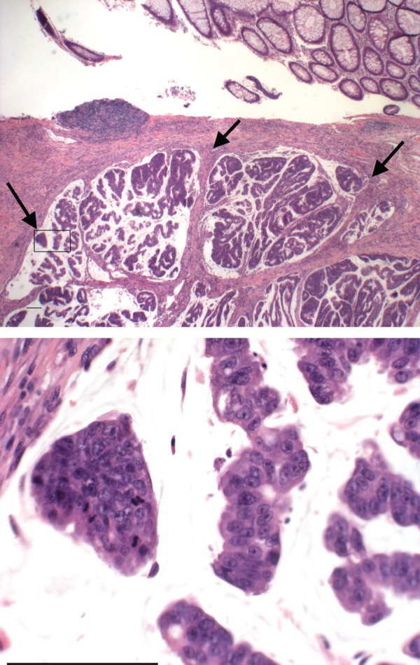
An example of the histological appearance of a human colon adenocarcinoma, along with adjacent normal colonic epithelium. The arrows point to sites of malignant cell invasion. See text for discussion. The area in the box on the low magnification image is enlarged in the lower panel to show mitoses, pleomorphic tumour nuclei and nucleoli. Scale bar, 100 µm. (Online version in colour.)