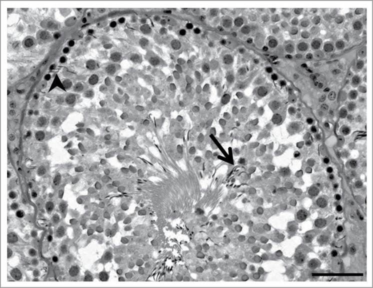
Figure 3.
Retained spermatids histopathology. In this rat stage X seminiferous tubule, step 19 spermatids are present in both the basal aspect (arrowhead) and apical aspect (arrow) of the seminiferous epithelium. In addition, this seminiferous tubule has a mottled appearance suggesting sloughing of the step 10 spermatid cohort may be occurring. Toxicant exposure in this example was a combination of 2,5-hexanedione and carbendazim, as described in the legend to Figure 1. Scale bar = 50 μm.

