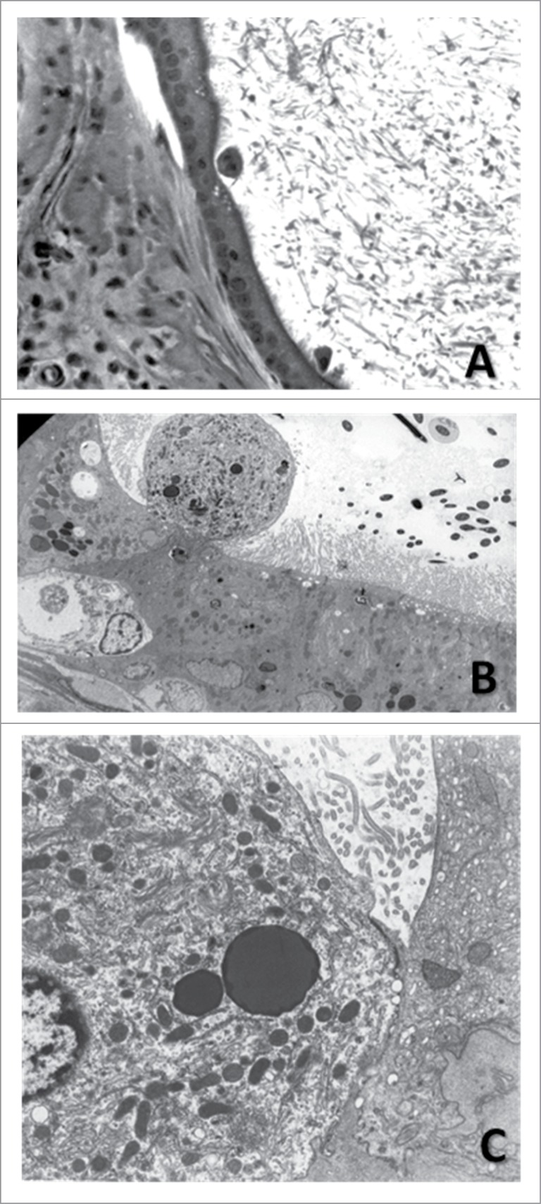
Figure 7.
Exfoliating principal cell in the cauda epididymidis of a hemicastrated rat treated with gossypol. (A) 20×, H&E. (B) Electron micrograph of the epididymal epithelium showing the exfoliation of a principal cell (2750×). (C) Cell indicated in B in greater detail. Notice the numerous mitochondria and lysosomes characteristic of principal cells (8000×). Adapted from Andrade et al. (2006).

