
Figure 1.
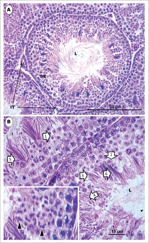
Japanese quail, Coturnix japonica. (A) represents an H&E-stained histological section of a transverse section of a seminiferous tubule surrounded by intertubular tissue in a sexually mature and active testis of the Japanese quail. L = lumen. (B) represents a higher power view of the seminiferous epithelium, which displays several spermatids and other germ cells. The epithelium shows several spermatids [1 to 6] at various steps of spermiogenesis, belonging to various cellular associations, but lying quite close to one another or appearing mixed, in some cases. Inset shows irregularly-shaped elongated spermatids at step 6 of spermiogenesis. L = lumen.