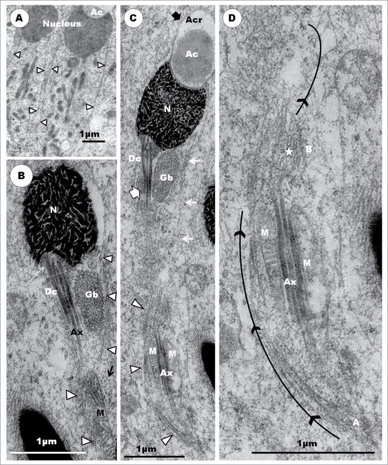
Figure 16.
(A) shows spermatids of step 7 displaying a well formed and complete microtubular sleeve (arrowheads) that extends nearly throughout the length of the spermatid. (B) and (C) show step 8 spermatids whose acrosomes twist a little out of line with the nuclei, and begin to exhibit convex surfaces which ultimately form the keels (thick arrow). Note the variable arrangement of the highly thickened strands of chromatin within the dumb-bell-shaped nuclei and the amorphous material (B), white broad arrow) which surrounds the distal centriole (Dc) as it attaches to the nucleus. The lumen of the distal centriole contains electron-dense linear but slightly irregular strands (white arrow), from which the singlet microtubules of the axoneme arise. In (B) and (C), the microtubular sleeve has moved toward the nucleus (white arrowheads in (B), and black arrows in (C)) [compare with Figs. 14F and 15A], and commences a rearrangement to form a loose bundle of microtubules (C) which is the precursor of the microtubular helix. (D) is an enlarged part of the lower half of (C), showing the profile of the microtubular helix in formation (curved black arrow), and transected roughly transversely at A and B (star). At point A, the bundle runs over the axoneme (Ax), but under it at point B. Note that the long strands of mitochondria (M) lie on all sides of the axoneme, as in Fig. F inset, and have, thus, not yet formed the mitochondrial helix. The granular body (GB) is elongated, but remains on only one side of the distal centriole (Dc) (B), (C).

