
Figure 4.
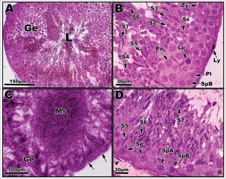
Light micrographs of seminiferous tubules of Graptemys pseudogeographica kohnii from September (A–B) and October (C–D). (A) Greatly enlarged seminiferous epithelium (Ge) dominated transforming spermatids; lumen (L) filled with mature sperm. (B) Segment of seminiferous epithelium reveals a type B spermatogonium (SpB), primary spermatocytes in various phases (Pl, Lp, Zy, Pa) previously mentioned in Figs. 2–3, a secondary spermatocyte (Ss), and morphological steps of spermatid development (S2-S7). A Leydig cell with lipid droplets (Ly) can be seen adhering to the basement membrane. (C) The height of seminiferous epithelium (Ge) is greatly reduced with concomitant increase of mature sperm (Ms) occupying the lumen; arrows point to nucleus of fibroblast cells. (D) Segment of seminiferous epithelium exhibiting a variety of germ cell types including type A and B spermatogonia and Sertoli cells (Se) along with spermatid steps 5–7. Arrowhead points to the nucleus of a fibroblast cell. Paraffin, (C); plastic, (A, B, and D).