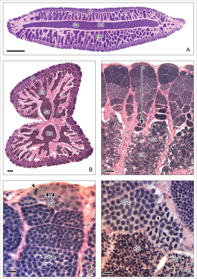
Figure 7 (See previous page).

Restricted lobular testis of the goodeid Goodea atripinnis. (A). Longitudinal section of the testis. (B). Transverse section of both testes. Spermatogenesis occurs at the periphery (P) or distal part of the testis lobules. Sperm are released, into a net of efferent ducts (Ed) which communicate with the central efferent duct (Cd). Efferent and central ducts are full of spermatozoa (Sz). (C). The lobules contain spermatocysts with synchronous germinal cells at different stages of spermatogenesis. Cysts at the later stages of spermatogenesis are located progressively closer (large arrow) to the efferent ducts (Ed) which contain abundant spermatozoa (Sz), organized in bundles called spermatozeugmata. Lobules do not have a central lumen. The interstitial tissue (It) is around the lobules. (D). Spermatogonia (Sg) are restricted to the periphery of the testis. A spermatogonium in mitotic metaphase is observed (arrow head). Cysts containing primary spermatocytes (1Sc) are seen. Interstitial tissue (It). (E). Cysts containing primary spermatocytes (1Sc), primary spermatocytes during metaphase of the first division of meiosis (arrow head), early spermatids (eSt) and late spermatids (lSt) are seen. Interstitial tissue (It). Hematoxylin-eosin. (A): Bar = 1mm; (B): Bar = 200 μm; (C): Bar = 50 μm; (D-E): Bars = 10 μm.
