Figure 5.

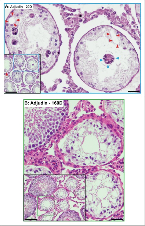
Phenotypes of the seminiferous epithelium in the testes of adult rats by day 20 (A) and day 160 (B) after treatment with a single dose of adjudin (50 mg/kg b.w., by oral gavage). (A) By day 20, tubules are virtually devoid of advanced germ cells. Only Sertoli cells and spermatogonia are found in the seminiferous epithelium. Occasionally, some multinucleate giant cells, such as multinucleate round spermatids are found (blue arrowheads). Vacuolization of seminiferous epithelium, illustrating Sertoli cell focal injury, is noted (red arrowheads). (B) By day 160, signs of rebounding of spermatogenesis is detected. As noted in the inset, at least 4 out of 10 tubules shown display signs of spermatogenesis with notable presence of elongating/elongated spermatids. Micrographs are magnified images from the corresponding boxed area shown in inset. Scale bars, 150 μm and 40 μm in insets.
