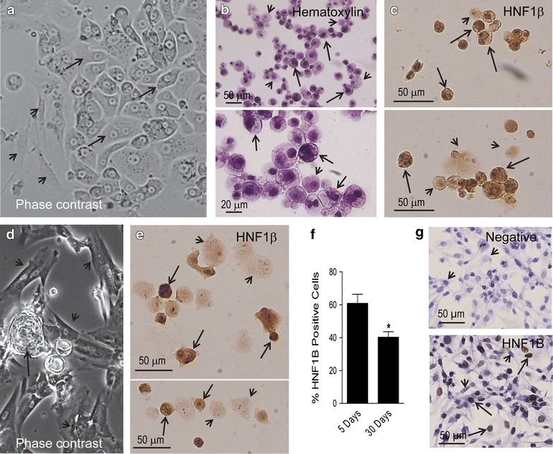
Fig. 3.

a Phase contrast image of a 5-day-old primary culture denoting a monolayer with an epithelial compartment depicting polygonal cells in a pavement-like arrangement (arrows) and a mesenchymal compartment (arrowheads) (original magnification ×20). b Cytospin cell preparations stained with hematoxylin and generated upon trypsinization of a section of a primary cell culture. The fields show two apparent types of cells of heterogeneous sizes, an abundant population of cells depicting dense chromatin and clear cytoplasm (arrows), and another population with less dense nuclei with prominent nucleolus, and less clear cytoplasm (arrowheads). c Cytospin cell preparations subjected to immunocytochemical staining for HNF1β. Many but not all cells show dark positive nuclei; arrows show positive-expressing cells whereas arrowheads show cells with negative expression; the panel also shows cells of different sizes. d Phase contrast image of a 30-day-old primary culture denoting higher proportion of cells with mesenchymal morphology (arrowheads)—when compared to a 5-day culture—always accompanied by epithelial-like cells growing in their vicinity (arrow) (original magnification ×10). e HNF1β staining in cytospin preparations of a 30-day-old primary culture displaying heterogeneity of expression, positive (arrows) or negative (arrowhead) cells. f Quantitation of the percentage of cells staining positive for HNF1β in cytospin preparations from 5-day or 30-day primary cultures of O-CCC. *p < 0.05 vs. 5 days (Student’s t-test). g Expression of HNF1β in a culture of SKOV-3 human cancer cells as counterstained with hematoxylin (upper panel negative; lower panel positive). Arrows depict positive immunostaining, whereas arrowheads indicate negative immunostaining
