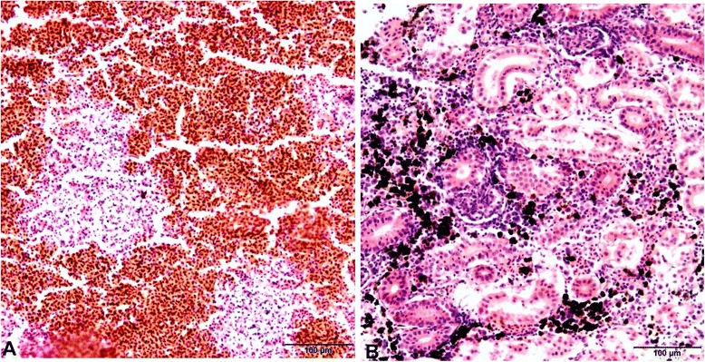
Figure 2.

Histological sections of spleen and kidney organs of rainbow trout infected with Y. ruckeri . A: multifocal necrosis can be seen in the spleen. B: degeneration of interstitial tissue and a marked increase in melano-macrophages can be seen in the kidney. Sections were stained with haematoxylin and eosin (H&E).
