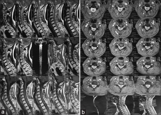
Figure 1.

Cervical magnetic resonance imaging 2 month after onset of herpes blisters. (a) Sagittal plane. MRIs were obtained in multiple images with different pulse sequences. Left paracentral-central protruded disk at C4-C5 and C5-C6 levels with compression to epidural space-left nerve root and stenosis of spinal canal and left neuroforamina were found. Central protruded disk was noted at C6-C7 level. Cervical magnetic resonance imaging 2 month after onset of herpes blisters. (b) Axial plane. MRIs were obtained in multiple images with different pulse sequences. Left paracentral-central protruded disk at C4-C5 and C5-C6 levels with compression to epidural space-left nerve root and stenosis of spinal canal and left neuroforamina were found. Central protruded disk was noted at C6-C7 level
