Figure 1.
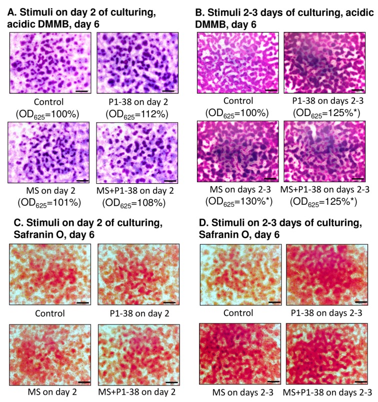
Effects of PACAP and/or mechanical load on matrix production of HDC. PACAP 1–38 at 100 nM was applied on day two of culturing (A,C) and continuously from day one (B,D). MS, mechanical stimulation applied on day two (A,C) and days 2–3 (B,D) for 30 min. (A,B) Metachromatic cartilage areas in six-day-old cultures were visualized with DMMB dissolved in 3% acetic acid. Metachromatic (purple) structures represent cartilaginous nodules formed by many cells and cartilage matrix rich in sulphated glycosaminoglycans (GAGs) and proteoglycans (PGs). Optical density (OD625) was determined in samples containing TB extracted with 8% HCl dissolved in absolute ethanol. Statistically significant difference of the extracted TB in cultures that received the loading regime and/or PACAP 1–38 vs. control cultures is marked by asterisk (* p < 0.05); (C,D) Safranin O staining for visualization of cartilage nodules of HDC. Original magnification was 4×. Scale bar, 500 µm. Representative data of three independent experiments are shown. (P1–38, PACAP 1–38; MS; mechanical stimulus).

