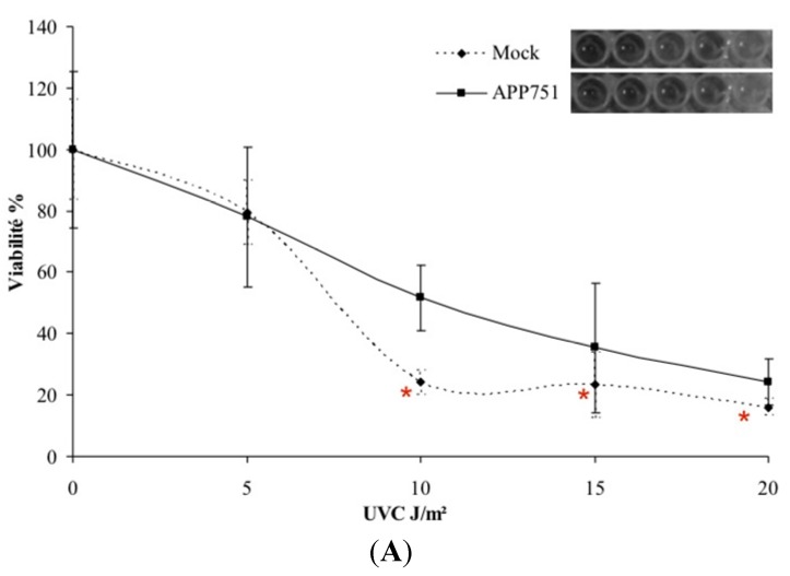
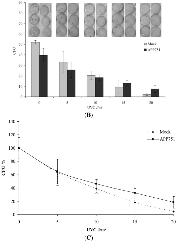
Figure 1.
UVC cytotoxicity at short and long-term. Mock and APP751-expressing cells were cultured 48 h prior to irradiation. Then, four doses of UVC 5, 10, 15 and 20 J/m2 were tested. For short-term cytotoxicity, cells were grown for additional 24 h and then the MTT assay was performed to assess cell viability (A). Significance was assessed through the Student t-test (n = 3). Data significantly different from mock cells (p < 0.05); For long-term toxicity, the cells were grown for an additional 12 days and then the colonies were revealed using crystal violet, and manually counted. The bar graph represents the number of colony forming units (CFU) in relation to the dose used; a representative picture of petri dishes following revelation with crystal violet is given above each bar of the graph, corresponding to each tested condition in triplicate (B); (C) represents the evolution of CFU in percentage Significance assessed through the Student t-test (n = 3).


