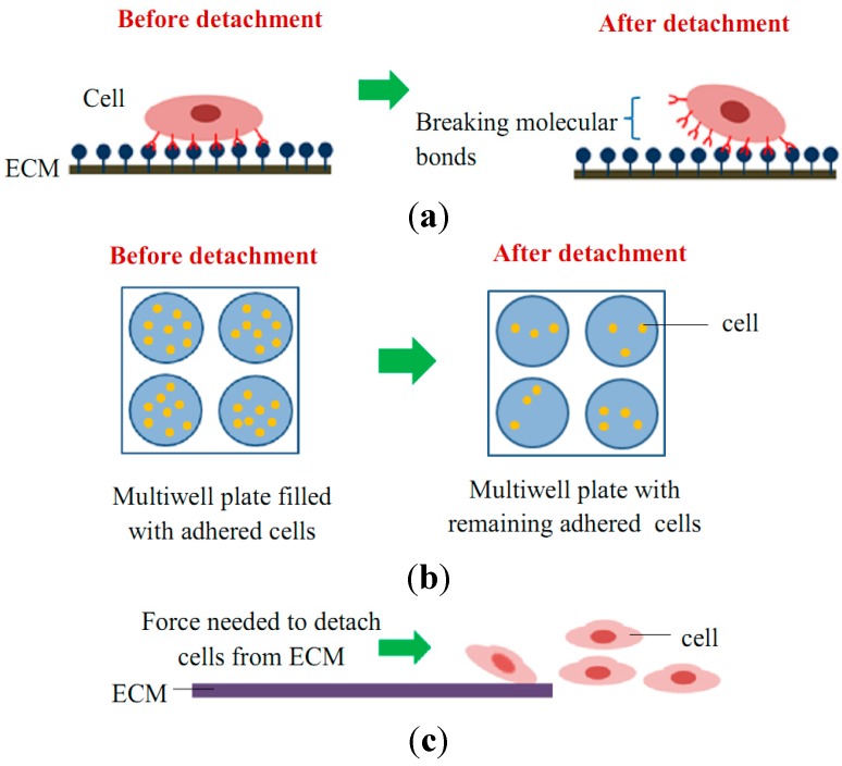
Figure 4.
Schematic diagram of cell adhesion detachment events for (a) single cell studies via the breakage of molecular bonds (e.g., SCFS, micropipette aspiration, and optical tweezer techniques); (b) cell population studies via static adhesion (e.g., centrifugation technique); and (c) cell population studies via dynamic adhesion (e.g., spinning disk, flow chamber, and microfluidic techniques).

