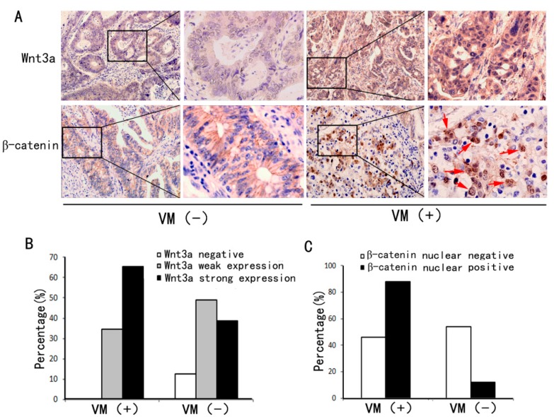
Figure 2.
Expressions of Wnt3a and β-catenin in the VM-positive and VM-negative groups. (A) Wnt3a expression was higher in VM-positive colon cancer tissue sections (right) than in VM-negative samples (left). In VM-positive sections, the tumor cells displayed nuclear β-catenin accumulation (red arrows), whereas those in the VM-negative section showed only membranous localization of β-catenin (immunohistochemical staining, ×200); (B) Percentages of Wnt3a negative, weak, and strong expression in the VM-positive and VM-negative groups; (C) Percentages of β-catenin nuclear positive and negative expression in the VM-positive and VM-negative groups.

