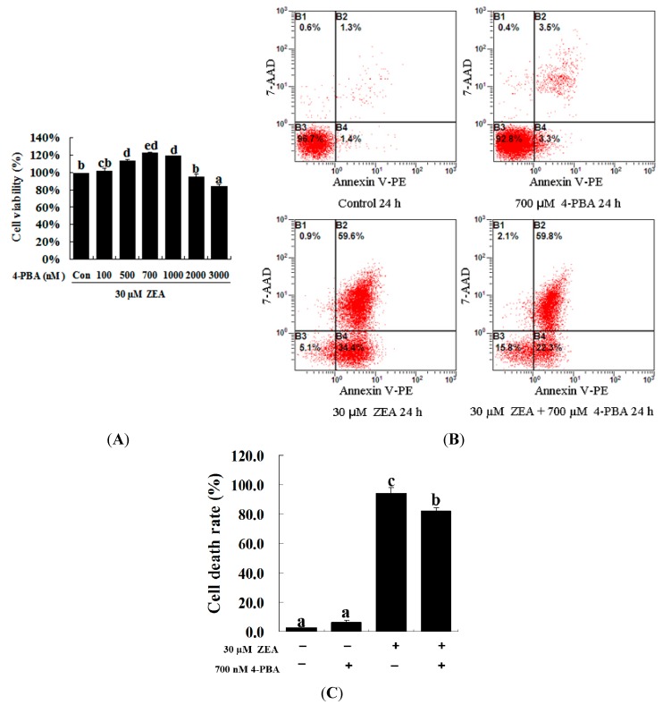
Figure 3.
Effect of 4-PBA on the growth of ZEA-treated RAW 264.7 macrophages. (A) RAW 264.7 macrophages were treated with 30 µM ZEA in the presence or absence of 4-PBA for 24 h. Different doses of 4-PBA (0–3000 nM) were used to assess concentration effects on cell viability, and cells were then processed for the MTT assay; (B,C) Apoptosis analysis was detected via flow cytometry. After exposure to 30 µM ZEA in the presence or absence of 700 nM 4-PBA for 24 h, RAW 264.7 macrophages were collected for Annexin V-PE/7-AAD staining followed by flow cytometric analysis. Statistical analysis of the cell death is shown in the bar graphs. Data are presented as the mean ± SEM of three independent experiments. Bars with different letters are significantly different (p < 0.05).

