Figure 4.
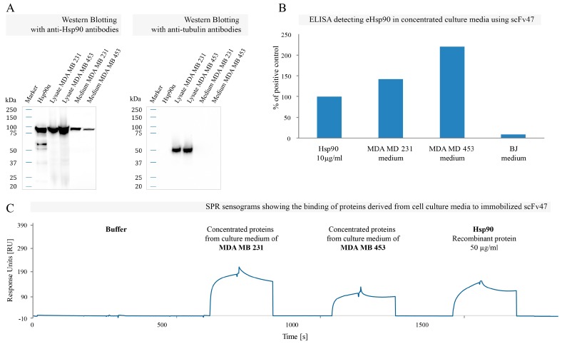
Interaction of scFv47 with eHsp90: (A) Western blot analysis of concentrated serum-free media derived from MDA MB 231 and MDA MB 453 cell cultures using specific anti-Hsp90 antibodies. Cell lysates containing cytoplasmic Hsp90 and recombinant Hsp90α served as positive controls; (B) The representative ELISA using scFv47 to detect eHsp90 in concentrated culture media derived from MDA MB 231, MDA MB 453 and BJ cells. The plate was coated with scFv47 and cell medium was applied. Detection of bound Hsp90 was performed using commercially available mouse anti-Hsp90 followed by anti-mouse-HRP antibody. Absorbance value from the controls with no antigen was subtracted from the obtained results, which were subsequently normalized as a fraction of absorbance from the wells where 10 µg/mL of recombinant Hsp90α was loaded (positive control); (C) The SPR sensograms showing the binding of Hsp90α to immobilized scFv47. After 38 hours of culturing MDA MB 231 and MDA MB 453 cells (in serum-free DMEM) media were collected, concentrated 100 times, buffer exchanged and injected on a sensor chip coated with ca. 1500 RU scFv47. Fifty micrograms per milliliters of the recombinant Hsp90α was injected as a positive control.

