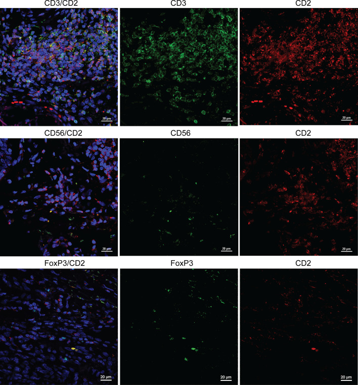
Fig. 3.
Co-localization of CD2/CD3 is more prevalent than CD2/CD56 and CD2/FoxP3 in melanoma specimens. In the top row, significant overlap of CD3+ (FITC) and CD2+ (Texas Red) cells was shown by immunofluorescence. In the middle row, expression patterns of CD56 (FITC) and CD2 (Texas Red) were dissimilar. In the bottom row, approximately 10 % of CD3+ (FITC, surface) cells express FoxP3 (Texas Red, intracellular). The results are representative of staining in three slides with high CD2 counts and three with low CD2 counts

