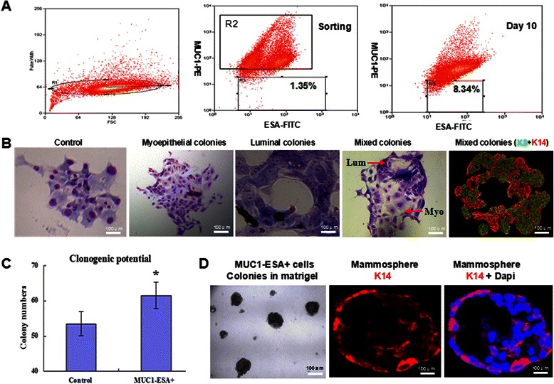
Fig. 1.

Tumor-initiation ability of MUC1−ESA+ cells sorted from mammary epithelium. a. MUC1−ESA+ subpopulation accounts for 1.35 % of MCF-10A cells when being sorted, and 8.34 % in serum-free culture on day 10. b. In the 2-D culture, sorted MUC1−ESA+ cells show three types of colonies including myoepithelial, luminal and mixed colonies, while the control cells (MCF-10A excluding MUC1−ESA+) displayed a unique type of colonies. c. The number of colonies and histogram of panel B (*, compared with the control, n = 6, P < 0.05). d. In the 3-D matrigel culture, sorted MUC1−ESA+ cells proliferate into colonies with duct-like structures and myoepithelial marker K14-DyLight 594 expression
