
Figure 1. Bioluminescence imaging in tissue culture and an animal model.
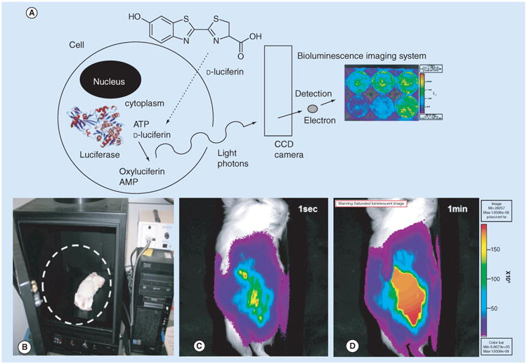
(A) Diagram of a single cell expressing FLuc, which enzymatically acts on d-Luciferin (substrate) to produce oxyluciferin + AMP and a photon of light. The photons are detected by the CCD camera, which produces the image, example shows tissue culture plate with FLuc expression virus. (B) IVIS-50 imaging system (Xenogen/Perkin Elmer., CA, USA) and BLI of FLuc expression from recombinant guinea pig CMV (GPCMV) in infected guinea pig pup. (C) The overlay image of the bioluminescence detected from the Fluc catalyzing D-Luciferin after 1 s and (D) 1 min onto image of the subject guinea pig pup.
CCD: Charge-coupled device.