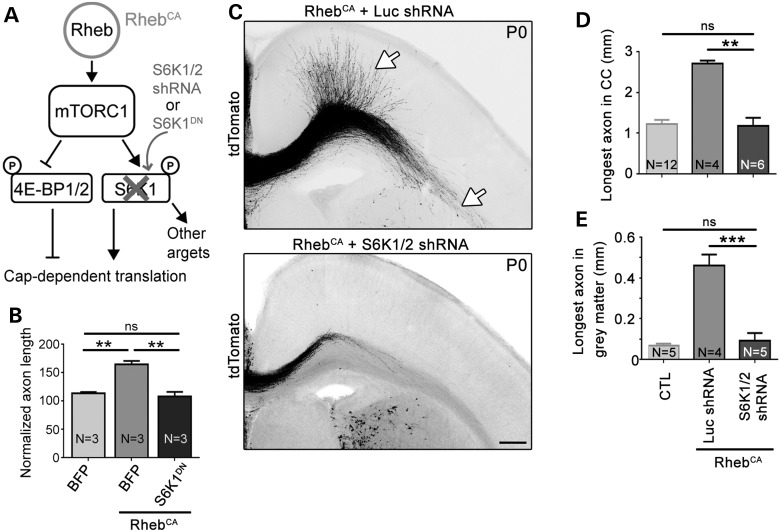
Figure 4.
Knocking down S6K1/2 prevents accelerated axon growth induced by hyperactive mTORC1. (A) Diagram depicting Rheb-mTORC1 pathway impinging on cap-dependent translation through 4E-BPs and S6Ks. (B) Bar graph of axon length (μm) in cultured cortical neurons nucleofected with BFP, S6K1DN with and without RhebCA. N = number of neuronal cultures. **P < 0.01 and ns: not significant with one-way ANOVA and Tukey post hoc test. The control condition is the same as that shown in Figure 2B. We measured >29 axons per culture and a mean of 197 and 153 axons in the BFP + RhebCA and S6K1DN + RhebCA conditions, respectively, using three sets of culture per condition. (C) Confocal images of tdTomato-fluorescent axonal projections from ACC neurons electroporated at E15 with RhebCA + luciferase (Luc) shRNA or RhebCA + S6K1/2 shRNA. The white arrows point to longer axons in gray and white matter in the RhebCA condition. Scale bar: 200 μm. (D and E) Bar graphs of the longest axon length in the corpus callosum (CC, in D, n = 10 axons in RhebCA + Luc shRNA and n = 11 axons in RhebCA + S6K1/2 shRNA, N = 4 mice each) and gray matter (in E, n = 10 axons in RhebCA + Luc shRNA and n = 11 axons in RhebCA + S6K1/2 shRNA, N = 4 mice each). Data for the control (CTL, BFP or GFP electroporation only) are those from Figure 1. N = the number of mice per condition indicated in each column. **P < 0.01 and ***P < 0.001 with one-way ANOVA and Tukey post hoc test.

