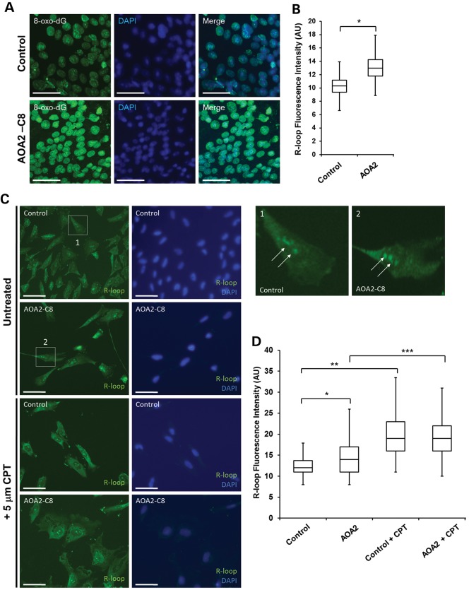
Figure 2.
Oxidative stress and R-loop formation in AOA2 iPSC. (A) Increased levels of oxidative DNA damage in AOA2 iPSC revealed by anti-8-oxo-dG immunostaining. Nuclei were stained with DAPI. Scale bar, 50 µm. (B) Quantitation of 8-oxo-dG fluorescence intensity (AU, arbitrary units) in both control and AOA2 iPSC. Fluorescence intensity was measured for 100 cells per experiment from three technical replicates from one experiment (*P < 0.05, Student's t-test). (C) Detection of R-loop in control and AOA2 iPSC using the S9.6 antibody in untreated (basal levels) and CPT-treated cells. DAPI counterstained nuclei. Scale bar, 20 µm. (D) Quantitation of average R-loop fluorescence intensity (AU) in untreated and CPT-treated iPSc. *, ** and *** indicate P < 0.001, one-way ANOVA.

