
Figure 1.
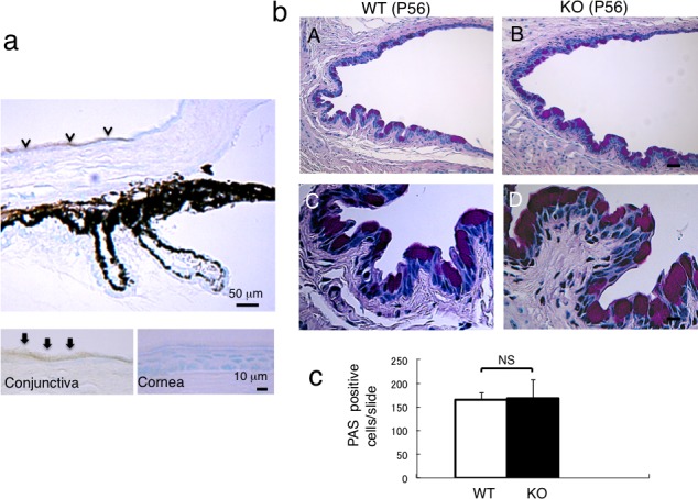
Expression of Muc16 in mouse ocular tissues and the histology of conjunctiva of WT and KO tissues. (a) Mucin 16 is expressed in conjunctival epithelium, but not in corneal epithelium. Higher magnification picture (insert) shows Muc16 in the superficial layer of the conjunctival epithelial cells. (b) Histology by PAS staining shows no obvious difference in the morphology of the conjunctival epithelium between WT (A) and KO (B) mouse. Scale bar: 50 μm. Higher magnification observation did not indicate differences in the morphology of goblet cells between WT (C) and KO (D) mouse. Scale bar: 10 μm. (c) The number of goblet cells was not affected by the loss of Muc16.