
Figure 3.
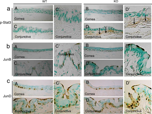
Expression pattern of phospho-Stat3 and AP-1 components in ocular surface. (a) Phospho-Stat3 was detected in subepithelial fibroblasts (arrows) in the KO conjunctiva ([D], higher magnification in [D']), but was not seen in the WT tissue ([C], higher magnification in [C']), as well as in the corneal epithelium and keratocytes in both genotypes of mice (A, B). (b) JunB protein was observed in some of the basal cells of WT and KO corneal epithelia (A, B) as well as in WT conjunctival basal epithelial cells ([C], higher magnification in [C']), while it was detected in the superficial epithelial cells in KO conjunctiva ([D], higher magnification in [D']). (c) Nuclear JunD is restricted to the basal layer cells of both the cornea and conjunctiva in both genotypes of mice (A–D). Scale bar: 10 μm.