
Figure 4.
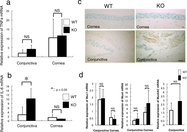
Expression of inflammatory components and membrane-associated mucin family members in cornea and conjunctiva. (a) Real-time reverse PCR clearly shows that there is no statistical difference in the expression level of TNFα mRNA between WT and KO mice in conjunctiva or cornea. (b) The loss of Muc16 promotes the mRNA expression of IL-6 in the conjunctiva (*P < 0.05), but not in the cornea. (c) Immunohistochemistry for IL-6 suggests that the subconjunctival tissue, but not the cornea, is the main source of IL-6 in the ocular surface in both genotypes of mice, and that IL-6 immunoreactivity seems more intense in the KO tissue compared with the WT one. Scale bar: 10 μm (cornea); 50 μm (conjunctiva). (d) The mRNA expression of Muc1 or Muc4 is higher in the conjunctiva or cornea, respectively, but there is no statistical difference in their expression levels between WT and KO tissues. The expression of Muc5AC mRNA in the conjunctiva is not significantly affected by the loss of Muc16.