
Figure 9.
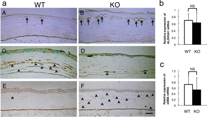
Immunohistochemical detection of macrophages and analysis of keratocyte phenotype in the healed cornea at 30 hours post debridement. (a) Immunohistochemistry detects F4/80-labeled macrophages, ALDH3A1 (keratocyte marker), and αSMA (myofibroblast marker). F4/80-labeled macrophages were more frequently observed beneath the regenerated epithelium in the KO mouse (B) compared with the WT cornea (A). The cells in the posterior stroma were labeled for ALDH3A1, while fibroblastic cells in the anterior stroma were not labeled in the WT cornea (C). In the KO cornea, the majority of the cells in the stroma were negative for ALDH3A1 (D). A few α-smooth muscle actin–positive myofibroblasts were detected in the WT stroma (E). Almost all the stromal cells were labeled with anti-αSMA antibody and thus were myofibroblasts in the KO cornea (F). Scale bar: 50 μm. Real-time PCR indicated no difference in the mRNA expression level of ALDH3A1 (b) and αSMA (c).