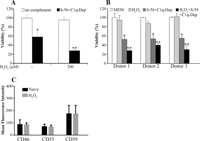
Figure 4.
Oxidative stress synergizes with complement to enhance cell death. (A) The ARPE-19 cells were pretreated with the indicated concentrations of H2O2 for 1.5 hours, primed with S-58 (1.2 mg/mL), and then incubated with 6% C1q-Dep. Cell viability was determined by WST-1 assay. *(P < 0.05) versus medium alone or oxidant alone. **(P < 0.05) versus complement alone or oxidant alone. (B) Retinal pigment epithelial cells from three donors were stimulated with H2O2 (200 μM) for 1.5 hours, primed with S-58 (1.2 mg/mL), and then incubated with 6% C1q-Dep. Cell viability was examined by WST-1 assay. *(P < 0.05) versus MEM alone or oxidant alone. **(P < 0.05) versus complement alone or oxidant alone. (C) The ARPE-19 cells were treated for 1.5 hours with H2O2 (0.5 mM). Cell surface mCRP levels were determined by FACS.

