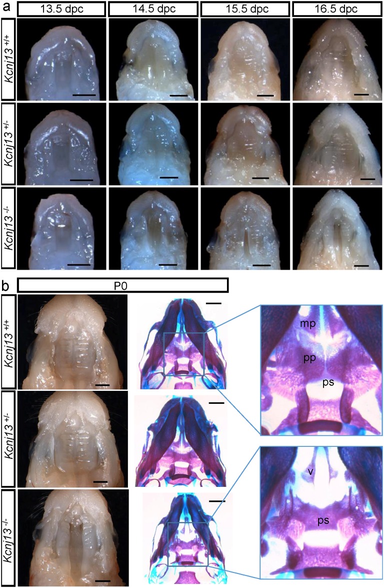
Fig 5. Palate formation during embryonic development in Kcnj13 +/+, Kcnj13 +/- and Kcnj13 -/- mice.
a. Dissected midfacial segments (without brain, mandible and tongue) from mouse heads collected from embryos 13.5–16.5 dpc. b. Mouse heads on postnatal day (P0) with dissected midfacial segments (left) and alizarin red and alcian blue skeletal staining (right). The palatine (pp) and maxillary (mp) processes are indicated In the Kcnj13 -/- specimen both the presphenoid (ps) and the vomer (v) are visible. Bar, 1 mm.

