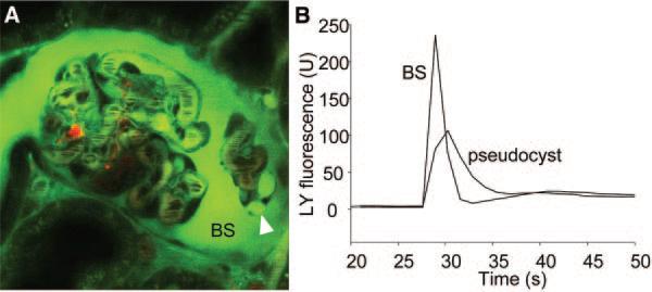
Figure 3.
Podocyte pseudocysts that form as a result of PAN treatment are enlargements of the subpodocyte space. (A) In vivo multiphoton image of an intact glomerulus from a PAN-treated Munich-Wistar-Fromter rat. The intravascular space (plasma) is labeled red using 70-kD dextran-rhodamine. (B) Lucifer yellow was injected into the carotid artery in bolus, and the time-lapse of its fluorescence (green) was recorded in the Bowman's space (BS) and inside pseudocysts (arrowhead) as shown in A. Lucifer yellow appeared and cleared from the BS quickly, whereas the filling and emptying in pseudocysts were slightly slower and delayed.

