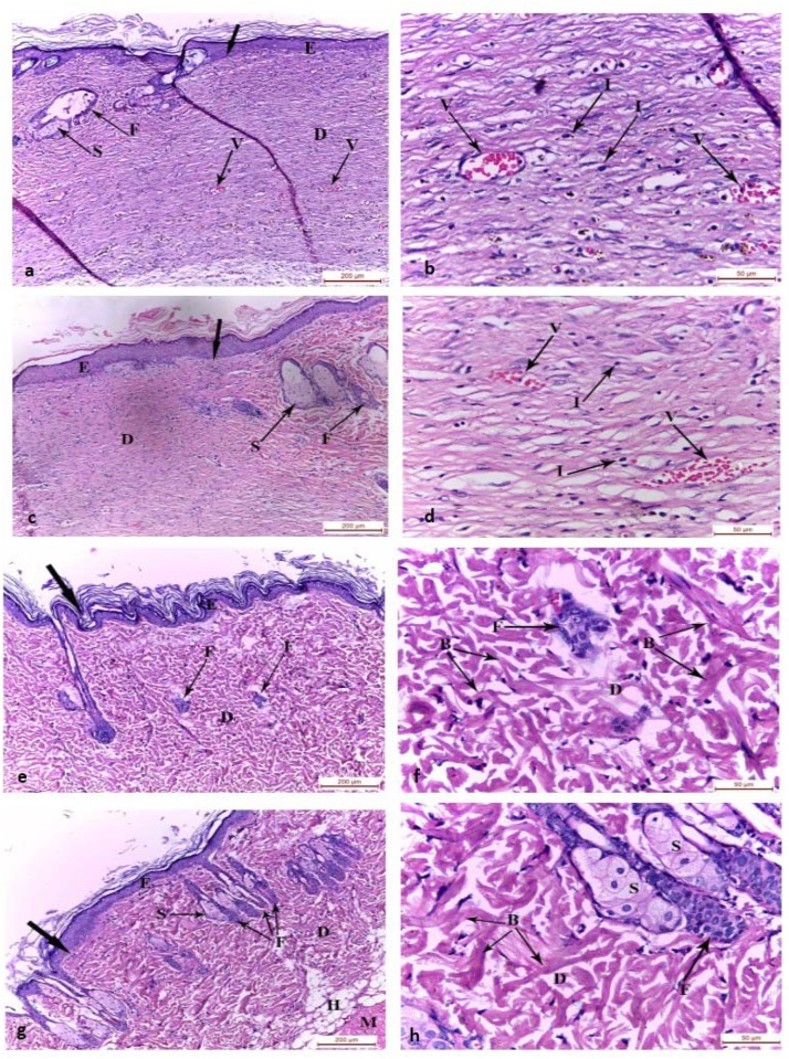
Fig 2. Photomicrographs of the skin wound area on the 15th day.
a,b) control group showing the wound area covered by regenerated epidermis. The regenerated dermis was filled with inflammatory cell infiltrates and many congested blood vessels with absence of hair follicles and sebaceous glands. The adjacent normal skin showed hair follicles and sebaceous gland. c,d) Group II showing the wound covered by regenerated epidermis on the granulation tissue in the regenerated dermis with absence of hair follicles and sebaceous glands. The adjacent normal skin showed hair follicles and sebaceous gland. Mild inflammatory cell infiltrates appeared in the dermis of the wound bed and congested blood vessels. e,f) Group III and g,h) Group IV wound area showing complete regeneration of the epidermis and dermis. The regenerated dermis showed fully regenerated hair follicles and sebaceous glands with absence of inflammatory cells and congested vessels. Connective tissue bundles were arranged in different directions. GT: granulation tissue, E: epidermis, D: dermis, H: hypodermis, M: muscle fibers, F: hair follicles, S: sebaceous gland, I: inflammatory cell infiltrate, B: collagen bundles, thick arrow: junction between the normal skin and the wound area, V: blood vessel, (H&E, a-c-e-g x100; b-d-f-h x400)

