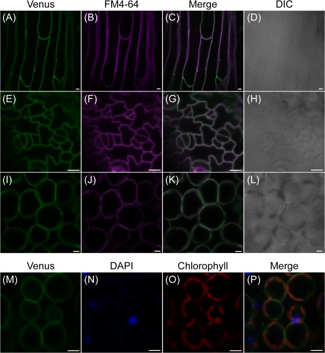
Fig 5. Subcellular localization of fluorescent AtPLC2-Venus in 12-day-old seedlings of ProPLC2:PLC2-Venus transgenic plants.
(A-D) Fluorescence of ProPLC2:PLC2-Venus (A) and staining of plasma membranes by FM4-64 dye (B) were merged (C) at stem epidermis. (E-H) Fluorescence of ProPLC2:PLC2-Venus (E) and staining of plasma membranes by FM4-64 dye (F) were merged (G) at leaf pavement and guard cells. (I-L) Fluorescence of ProPLC2:PLC2-Venus (I) and staining of plasma membranes by FM4-64 dye (J) were merged (K) at leaf mesophyll cells. (D), (H), and (L) are differential interference contrast (DIC) images for each sample. (M-P) Fluorescence of ProPLC2:PLC2-Venus (M), staining of nuclei by DAPI (N) and chlorophyll autofluorescence (O) were merged (P) at leaf mesophyll cells. Scale bars are 10 μm.

