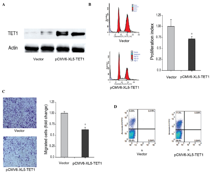
Figure 4.
Overexpressed TET1 inhibits ACHN cell proliferation and metastasis, and improves apoptosis. (A) TET1 was overexpressed in the ACHN renal carcinoma cell line. The ACHN cells were transfected with pCMV6-XL5-TET1 and screened using puromycin to generate a stable cell line. The protein levels in the whole cell extracts were analyzed using western blot analysis. (B) Proliferation rate of the TET1-overexpressed ACHN cells was determined using flow cytometric analysis. The PI was calculated as follows: PI= (S + G2/M) / (S + G2/M + G0/G1). (C) Metastatic ability of the TET1-overexpressed ACHN cells was determined using a Transwell assay. (D) Apoptotic rate of the TET1 overexpressed ACHN cells was analyzed using flow cytometry subsequent to labeling with PI and annexin V. (*P<0.05, compared with the vector group). TET1, ten-eleven translocation methylcytosine dioxygenase 1; FITC, fluorescein isothiocyanate.

