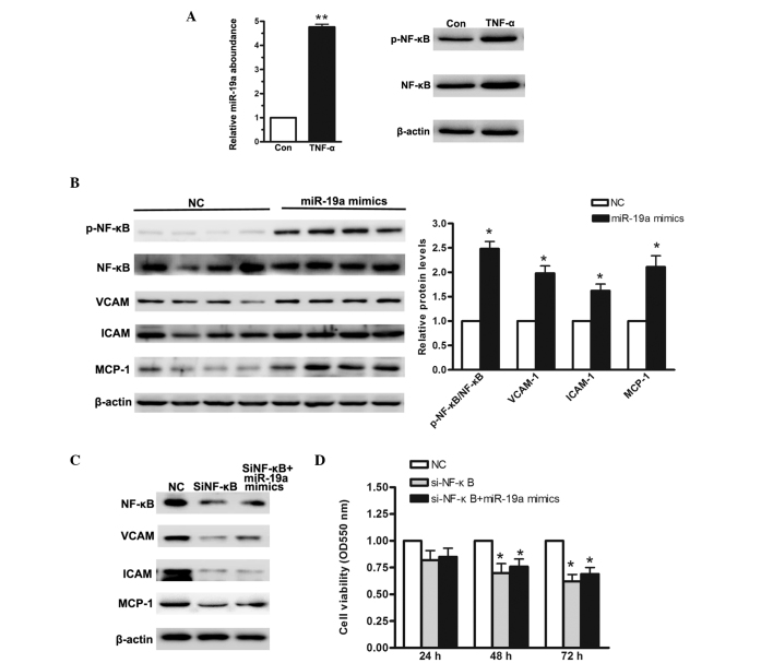
Figure 3.
NF-κB signaling pathway is activated when miR-19a is overexpressed in the MGC-803 human gastric carcinoma cell line. (A) Reverse transcription-quantitative polymerase chain reaction was used to determine the relative levels of miR-19a when MGC-803 cells were treated with 10 ng/μl TNF-α for 48 h. (B) Western blot analysis of NF-κB activation and its downstream regulators when miR-19a was overexpressed. (C) Western blot analysis of siRNA targeting NF-κB. (D) MTT assay demonstrated a low cellular proliferation rate in cells cotransfected with si-NF-κB and miR-19a mimics. Data are presented as the mean ± standard error of three independent experiments. *P<0.05, vs. control. TNF-α, tumor necrosis factor-α; NF-κB, nuclear factor-κB; VCAM, vascular cell adhesion molecule; ICAM, intercellular adhesion molecule; MCP, monocyte chemoattractant protein; miR, microRNA; si, small interfering; p-, phosphorylated; NC, negative control; Con, control.

