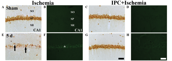
Figure 2.

NeuN immunohistochemistry (the first and third columns) and F–J B histofluorescence staining (the second and fourth columns) in the CA1 region of the ischemia group (left two columns) and IPC + ischemia group (right two columns) at (A–D) sham and (E–H) 5 days-post ischemia-reperfusion. In the sham group, several NeuN+ neurons and no F–J B+ cells were detected in the SP. In the ischemia group, a few NeuN+ neurons (black arrows) and several F–J B+ cells (white asterisks) were detected in the SP at 5 days post-ischemia. However, in the IPC + ischemia-group, abundant NeuN+ neurons and no F–J B+ cells were detected in the SP at 5 days post-ischemia. Scale bar=50 µm. NeuN, neuronal nuclei; F–J B, fluoro-Jade B; IPC, ischemic preconditioning; SP, stratum pyramidale; SO; stratum oriens, SR; stratum radiatum.
