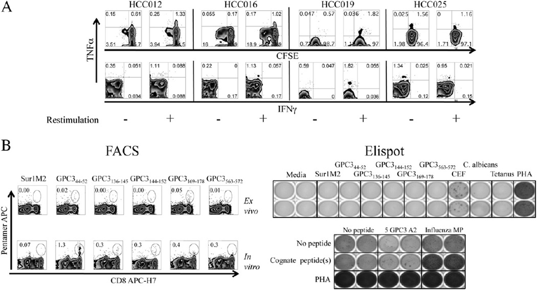
Figure 4.
Cytokine and degranulation profile of 15mer peptide short-term in vitro-expanded T-cells. A. Representative intracellular cytokine FACS plots showing CFSE dilution versus TNFα (upper) and TNFα vs IFNγ (lower) for four HCC patients with glypican-3-specific TNFα responses after 1 week of in vitro expansion using 15mer pooled peptides. B. Example in which PBMC from HLA-A2+ HCC patient were expanded with 9–10mer optimal peptides. Pentamer frequency ex vivo (top left) and after expansion in vitro (bottom left) for Sur1M2 and 5 GPC3 pentamers are shown. Ex vivo IFNγ Elispot (top right) shows no IFNγ+ responses against any glypican-3 peptide with lack of increase in IFNγ production by Elispot after restimulation with cognate antigen (bottom right).

