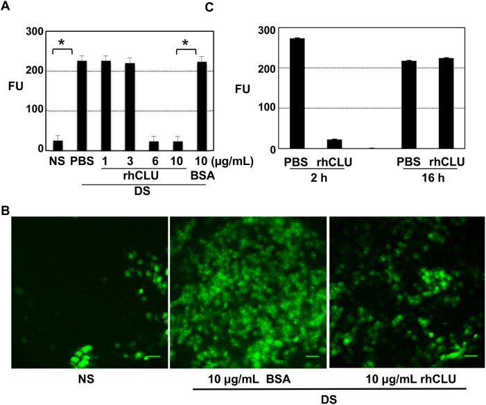
Fig 4. Topical CLU directly seals the ocular surface barrier disrupted by desiccating stress.
The standard desiccating stress (DS) protocol was applied for 5-days to create ocular surface disruption. Non-stressed (NS) mice housed under normal ambient conditions served as a baseline control. Eyes with desiccating stress were then treated topically, a single time, with 1 uL of CLU formulated in PBS, 1 uL of BSA formulated in PBS for comparison, or 1 uL of PBS control. Barrier disruption was assayed by measuring corneal epithelial uptake of fluorescein (FU = Fluorescence Units at 521 nm). Values are expressed as the mean ± SD. (A) Eyes were treated a single time with recombinant human CLU (rhCLU) at 1, 3, 6 or 10 ug/mL, BSA at 10 ug/mL, or PBS. Fifteen minutes later, the fluorescein uptake test was performed, before there was time for barrier repair to occur. *P<0.0001 (n = 4). (B) Images of central cornea from the experiment shown in (A), obtained using laser scanning confocal microscopy at 10X magnification. One representative image out of two independent experiments is shown. Scale bar = 100 um. (C) Eyes were treated a single time with recombinant human CLU (rhCLU) at 10 ug/mL (right eyes) or PBS (left eyes). Then the mice were kept further for 2 h or 16 h while continuing with the same desiccating stress protocol. The fluorescein uptake test was performed following the indicated time period to assess the time length of CLU treatment effect. *p<0.0001 (n = 4)

