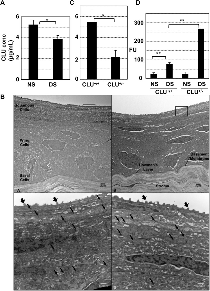
Fig 7. Causal association between endogenous CLU concentration in tears and ocular surface barrier vulnerability.
(A) Tears were collected from mice housed under normal ambient conditions or after application of the standard desiccating stress (DS) protocol for 5-days, and ELISA was used to measure CLU concentration (*P = 5x10⁻⁸ n = 6, student’s t-test). (B) Representative transmission electron microscopy comparing images of anterior cornea from wild type C57BL6/J mice (A and C) and mice with homozygous CLU-/- knockout on the C57BL6/J background (B and D). In low power (4000x) magnifications (A and B), five layers of epithelial cells divided into squamous, wing, and basal cell regions are visualized along with an intact basement membrane and Bowman's layer in both types of animals. Higher power images (C and D, 20,000x) of similar regions to those boxed in panels A and B show numerous surface microplicae (fat arrows) in both genotypes. Desmosomes (thin arrows) are similar in both frequency and structure. Higher power images (not shown) demonstrate intact adherens junctions in both genotypes. (C) Tears from wild type or heterozygous CLU+/- knockout mice kept at ambient conditions were collected and ELISA was used to measure CLU concentration (p = 2.1x10⁻⁵; n = 7, student’s t-test). (D) Wild type mice or heterozygous CLU+/- knockout mice were subjected to the standard desiccating stress protocol, but without scopolamine injection for four weeks and then ocular surface barrier integrity was measured by fluorescein uptake (**p<0.0001, n = 4).

