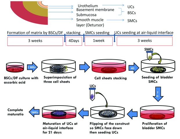
Fig. 1.
The study plan. In the upper panel, each cell type is extracted from a different portion within the same single bladder biopsy where urothelial cells (UCs) were extracted from mucosa, bladder stroma cells (BSCs) mainly from submucosa and smooth muscles (SMCs) from detrusor muscle. The main steps of urinary equivalent formation with time scale for each step is shown in middle panel. Illustrations for the steps of equivalent production with self-assembly technique are seen in the lower panel.

