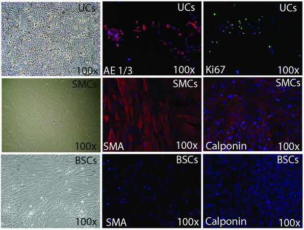
Fig. 2.
Phenotype characterization of 3 cell types extracted from bladder biopsy with immunofluorescence. Urothelial cells (UCs) had the characteristic appearance under phase contrast microscopy and their phenotype with phase contrast microscopy, expressed epithelial markers (AE1/AE3) with immunofluorescence. Smooth muscle cells (SMCs) exhibited SMCs markers; smooth muscle actin and calponin. Bladder stromal cells (BSCs) manifested weak expression for SMA and no expression for calponin.

