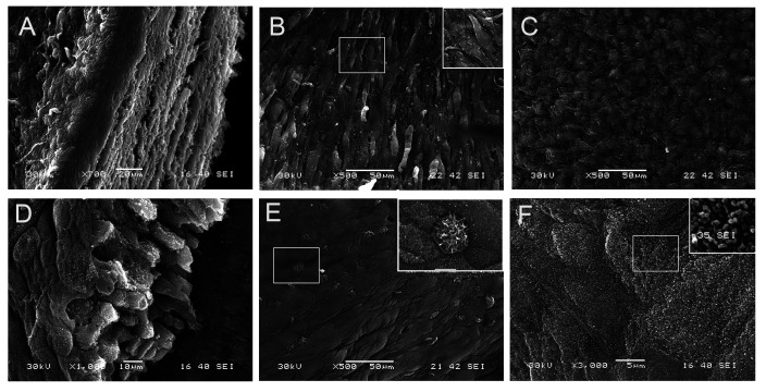
Fig. 7.
Scanning electron microscopy for bladder stroma cells- smooth muscle cells (SMCs) reconstructs. A: Cut section through the whole equivalent showing the different layers and thee thick stroma. B: SMC surface with aligned smooth muscle cells. The box in the upper right corner emphasizing the spindle shaped appearance of SMCs. C: The upper surface of the stroma with tightly packed collagen. D: Cut section though the urotheium with multiple layers resting on basement membrane (BM). E: the apical surface of urothelium formed of umbrella cells with well-defined borders and scattered microvillous cells (magnified picture in the upper right corner). F: the apical membrane of umbrella cells with uroplakins particles (magnified picture in the upper right corner).

