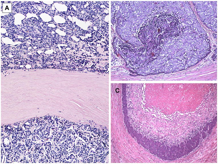
Fig. 3.
Conventional type A thymoma versus atypical type A thymoma variant. A. Conventional type A thymoma exhibiting areas composed of spindle cells (upper half) and polygonal cells (lower half) separated by a broad fibrouis septum. B. and C. Atypical type A thymoma variant showing various degrees of spontaneous necrosis in polygonal cell areas. This tumor invaded the lung and showed a missense mutation (chromosome 7 c.74146970T>A) in the GTF2I gene that is common in type A thymomas but rare in type B thymomas and thymic carcinomas.23

