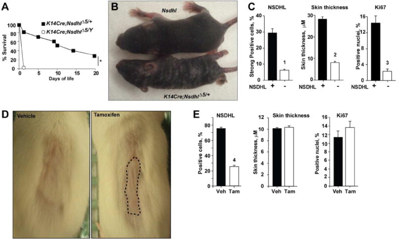
Figure 1.

Blockade of keratinocyte proliferation by conditional inactivation of NSDHL. (A) Survival of K14Cre;NsdhlΔ5/+ females and K14Cre;NsdhlΔ5/Y males. Shown is pooled data from 3 independent crosses; *, p=10−4. (B) Conditional inactivation of Nsdhl produces hyperkeratotic, scaly patches and a striped coat in 1 week old heterozygous K14Cre;NsdhlΔ5/+ female pups. (C) NSDHL-deficient areas of the dorsal skin in 4 week-old K14Cre;NsdhlΔ5/+ females show reduced thickness and proliferation of the basal layer keratinocytes (Ki67-positive nuclei). See also Figure S3. Digital imaging data are represented as mean±SEM. (D) Delayed hair regrowth (dashed line) following conditional Nsdhl ablation in the skin of adult K5CreERT;Nsdhlflx5/Y mice following oral administration of vehicle (left, corn oil) or tamoxifen (right, 40 mg/kg) for 5 consecutive days at age of 12 weeks. One week after treatment, the coat from the dorsal skin was depilated and hair regrowth was imaged 1 week after depilation. (E) No change in interfollicular epidermis Ki67-positive cells or epidermis thickness in tamoxifen-treated adult K5CreERT;Nsdhlflx5/Y mice. See also Figure S4. Digital imaging data are represented as mean±SEM, p-values: (1) p=3*10−6; (2) p=3*10−16; (3) p=2*10−5; (4) p= 2*10−16, based on Wilcoxon test.
