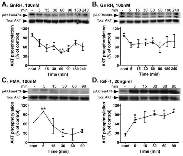
Figure 7.
Time course of AKT activity by GnRH, PMA and IGF-1 in αT3-1 cells. αT3-1 cells were serum-starved for 16 h, followed by stimulation with GnRH, PMA (100nM each) or IGF-1 (20ng/ml) for up to 240 min. After treatment cell lysates were analyzed for AKT activity by Western blotting using an antibody for phospho-AKT- Ser473 (pAKTser473) or phospho-AKT-Thr308 (pAKTthr308). Total AKT was detected with polyclonal antibody as a control for sample loading. A representative blot is shown and similar results were observed in three other experiments. The results from three experiments are shown as mean±S.E.M of control phosphorylation.**p-val≤0.01; *p-val≤0.05 vs. control.

