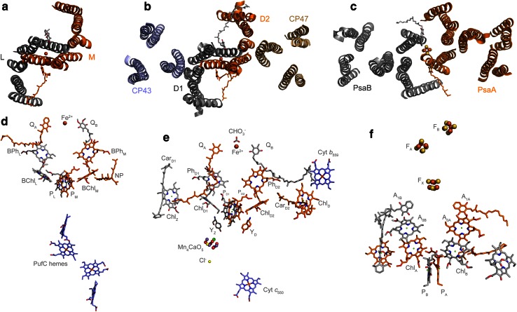
Fig. 1.
Reaction center architecture and cofactors. a Top view of a Type II reaction center from Blastochloris viridis highlighting the position of the transmembrane helices, PDB ID: 2PRC (Lancaster and Michel 1997). b Top view of Photosystem II from Thermosynechococcus vulcanus where only the transmembrane helices from the reaction center subunits (D1 and D2) and antenna proteins (CP43 and CP47) are highlighted, PDB ID: 3ARC (Umena et al. 2011). c Top view of Photosystem I from Synechococcus elongatus, PDB ID: 1JB0 (Jordan et al. 2001). d Cofactors in the Type II reaction center from B. viridis, the pigments colored in orange are coordinated or held by the M subunit, while those in gray by the L subunit. e Cofactors in Photosystem II, only those held by the D1 (gray) and D2 (orange) proteins are shown. f Cofactors in Photosystem I, those coordinated by the PsaA protein are colored in orange, and those by the PsaB are colored in gray. Besides the main pigments involved in charge separation, there are remarkable similarities in the position of certain accessory carotenoids and chlorophylls between Photosystem II (CarD1, CarD2, ChlZ, ChlD) and Photosystem I, suggesting that these may have been present in the ancestral reaction center

