Fig. 2.
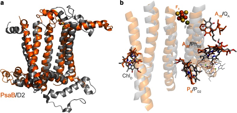
Comparison of a Type II reaction center subunit, D2 of Cyanobacterial Photosystem II (gray), and a Type I reaction center subunit (last five transmembrane helices) of the PsaB protein of Photosystem I (orange). a Overlap of D2 and PsaB; the F X cofactor from Photosystem I and the non-heme Fe2+ from Photosystem II have been displayed as spheres to show their relatives positions. b Overlap of some of the cofactors coordinated by D2 (gray) and PsaB (orange). The peripheral chlorophylls, ChlZ and ChlD, are conserved in these two subunits (Baymann et al. 2001). This histidine is also found in the sequences of all Type I reaction centers, with the exception of C. thermophilum and the anoxygenic Type II reaction centers. Its presence in Photosystem II implies that it was present in the most ancestral reaction center. The position of some of the chlorophylls, the quinones, F X, and non-heme Fe2+ is also very similar, yet the mode of coordination varies depending on the type of reaction center. The alignment of the subunits was made with the CEalign (Jia et al. 2004) plugging of Pymol (Molecular Graphics System, Version 1.5.0.4 Schrödinger, LLC)

